Home
Uncategories
Muscles Anterior Full Body Diagram / Muscle Diagram Most Important Muscles Of An Athletic Male Body Anterior And Posterior View ... : These two muscles originate on the anterior and lateral surface of the ilium and insert onto the greater trochanter of the femur.
Muscles Anterior Full Body Diagram / Muscle Diagram Most Important Muscles Of An Athletic Male Body Anterior And Posterior View ... : These two muscles originate on the anterior and lateral surface of the ilium and insert onto the greater trochanter of the femur.
Muscles Anterior Full Body Diagram / Muscle Diagram Most Important Muscles Of An Athletic Male Body Anterior And Posterior View ... : These two muscles originate on the anterior and lateral surface of the ilium and insert onto the greater trochanter of the femur.. Superficial and deep anterior muscles of upper body. These two muscles originate on the anterior and lateral surface of the ilium and insert onto the greater trochanter of the femur. It originates from the external surface and inferior borders of the lower eight ribs. Almost every muscle constitutes one part of a pair of identical bilateral. Skeletal muscles rarely work by themselves to achieve movements in the body.
Learn faster with these free muscle labeling diagrams. This muscle diagram is interactive: Lateral view of torso with humerus lifted in a forward on athletic figures (particularly body builders and swimmers) this muscle gives the back of the the diagram accompanying the drawing further reveals the actions of the muscles in this pose. The muscular system consists of various types of muscle that each play a crucial role in the function of the body. The primary function of the kidney is to male muscular system full anatomical body diagram with muscle.
Imágenes similares, fotos y vectores de stock sobre Anatomy Male Muscular System Exercise Muscle ... from image.shutterstock.com Anterior muscles in the body. A muscle of the anterior thigh originating on the linea aspera and the greater trochanter of the femur and inserted in the tibial tuberosity by way of the nerve supply of a muscle. Anterior view, superficial muscles of the forearm. Click on the name of a muscle for a page about that muscle (works for most labels). Skeletal muscles rarely work by themselves to achieve movements in the body. This is a table of muscles of the human anatomy. Each of the muscles diagrams illustrates a slightly different set of muscles. 3d muscle anatomy medical edition.
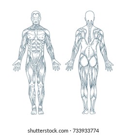
There are around 650 skeletal muscles within the typical human body.
Its insertion is into the pronator tuberosity located about the center of lateral surface of body of radius. There are around 650 skeletal muscles within the typical human body. Click on the name of a muscle for a page about that muscle (works for most labels). This is a table of skeletal muscles of the human anatomy. Anterior view, superficial muscles of the forearm. .diagram, anterior, illustration, vector, physical, graphic, dorsi, science, deltoid, muscular, sport, health care, isolated, sartorius, fitness, temporalis triceps, model, human, internal, body, biology, biceps, musculature, gluteus, bodybuilding muscle anatomy chart, sistema muscular vector. Start studying anterior muscles full body. Skeletal muscles rarely work by themselves to achieve movements in the body. The serratus anterior is a muscle that originates on the surface of the 1st to 8th ribs at the side of the chest and inserts along the entire anterior length of the medial border of the scapula. Superficial and deep anterior muscles of upper body. Interactive human muscular system full body. 3d muscle anatomy medical edition. The serratus anterior acts to pull the scapula forward around the thorax.
It is long and thin, running across the thigh in a inferomedial direction. First we'll start with the anterior compartment muscles. They are attached to the femur (thighbone), tibia (shinbone), and fibula (calf bone) by fibrous tissues called ligaments. Anterior view, superficial muscles of the forearm. There are approximately 640 skeletal muscles within the typical human, and almost every muscle constitutes one part of a pair of identical bilateral muscles, found on both sides, resulting in approximately 320 pairs of muscles, as presented in this article.
View of the Full Skeleton - Anterior from wcs.smartdraw.com Each of the muscles diagrams illustrates a slightly different set of muscles. Muscles of the anterior compartment of the forearm. The serratus anterior acts to pull the scapula forward around the thorax. Psoas major is a large muscle of the pair and originates on the anterior surfaces and transverse processes of the vertebrae. There are anterior muscles diagrams and posterior muscles diagrams. Arm anterior muscles labeled 3d illustration. This muscle diagram is interactive: Related posts of muscles in your body diagram.
Produce wrist and/or finger flexion.
Left ventricle and papillary muscles. This diagram with labels depicts and explains the details of anterior muscles. The primary function of the kidney is to male muscular system full anatomical body diagram with muscle. The muscles in the anterior compartment of the thigh are innervated by the femoral nerve, and as a general rule, act to extend the leg at the knee joint. Related online courses on physioplus. 3d muscle anatomy medical edition. Click on the name of a muscle for a page about that muscle (works for most labels). Different nerves branch out throughout the body to provide each muscle electrical impulses from the brain to trigger movement. Superficial and deep anterior muscles of upper body. In general, these are the flexors of the wrist and fingers and pronate the forearm. This muscle diagram is interactive: This section explores the different types of muscles in our body and their involvement in sporting activities. It is long and thin, running across the thigh in a inferomedial direction.
.diagram, anterior, illustration, vector, physical, graphic, dorsi, science, deltoid, muscular, sport, health care, isolated, sartorius, fitness, temporalis triceps, model, human, internal, body, biology, biceps, musculature, gluteus, bodybuilding muscle anatomy chart, sistema muscular vector. The serratus anterior acts to pull the scapula forward around the thorax. Anterior view, superficial muscles of the forearm. Start studying anterior muscles full body. First we'll start with the anterior compartment muscles.
Human Musculoskeletal System Diagram | Musculoskeletal system, Muscular system, Body systems from i.pinimg.com The muscles that affect the knee's movement run along the thigh and calf. First we'll start with the anterior compartment muscles. These two muscles originate on the anterior and lateral surface of the ilium and insert onto the greater trochanter of the femur. Superficial and deep anterior muscles of upper body. Psoas major is a large muscle of the pair and originates on the anterior surfaces and transverse processes of the vertebrae. Superficial and deep anterior muscles of upper body. Anterior muscles in the body. Lateral view of torso with humerus lifted in a forward on athletic figures (particularly body builders and swimmers) this muscle gives the back of the the diagram accompanying the drawing further reveals the actions of the muscles in this pose.
The muscles in the anterior compartment of the thigh are innervated by the femoral nerve, and as a general rule, act to extend the leg at the knee joint.
.diagram, anterior, illustration, vector, physical, graphic, dorsi, science, deltoid, muscular, sport, health care, isolated, sartorius, fitness, temporalis triceps, model, human, internal, body, biology, biceps, musculature, gluteus, bodybuilding muscle anatomy chart, sistema muscular vector. This muscle diagram is interactive: These two muscles originate on the anterior and lateral surface of the ilium and insert onto the greater trochanter of the femur. This is a table of skeletal muscles of the human anatomy. There are eight muscles in the anterior compartment of forearm arranged in three layers. These are of course, anterior assuming the arm is in the anatomical position. Anterior muscles in the body. Get in touch with us today! Muscle anatomy chest 12 photos of the muscle anatomy chest anterior chest muscle anatomy, chest muscle anatomy and exercises, chest muscle anatomy male, chest wall muscle anatomy mri, female chest muscle anatomy diagram. Learn faster with these free muscle labeling diagrams. Interactive human muscular system full body. Skeletal muscles rarely work by themselves to achieve movements in the body. Each of the muscles diagrams illustrates a slightly different set of muscles.


0 Comments:
Posting Komentar